X-ray imaging |
| X-ray imaging technique | |||||
|
|
|||||
|
|
|||||
|
|
|||||
| X-ray imaging of biomedical application | |||||
|
|
|||||
|
|
|||||
|
|
|||||
| X-ray imaging technique
|
X-ray grating interferometer for biological imaging

|
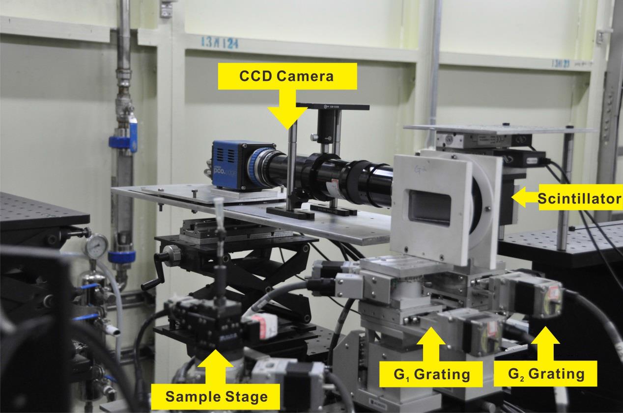
Together with collaborators, we has built a grating-based phase-contrast (GPC) imaging system at BL13W1 beamline,SSRF (Shanghai Synchrotron Radiation Facility). The setup has comparable density and spatial resolutions as compared with similar setups at some leading synchrotron facilities around the world. In applying the setup for biomedical applications, we has established close collaborations with medical researchers. Exploiting the high density and spatial resolutions of the imaging setup, we has investigated the micro-angiogenesis process in tumor development and the non-invasive identification of metastases at early stage. The work has been published on J.Synchrotron Rad,19, 821-826(2012). ( PDF ) |
Ultrafast X-ray imaging technique

|
While working at Argonne National Laboratory in Chicago, together with collaborators, we developed the ultrafast synchrotron X-ray imaging system. The imaging speed reached 300,000 frames per second and an exposure time of 150 picoseconds per frame. The new imaging technique has shown very important applications in many disciplines, such as the optically opaque dense high-speed engine sprays. Also it shows great potential for fluid imaging owing to the edge-enhancement, penetrative and immune of optical distortion properties(Phys. Rev. Lett. 100, 154502, (2008);Phys. Rev. Lett. 100, 104501, (2008)). The work has been published on Nature Physics, 4, 305–309 (2008). ( PDF ) |
Sagittally focusing double-multilayer monochromator

|
Ordinary X-ray crystal monochromator has very narrow energy bandwidth, and the corresponding full-field imaging speed is on the order of seconds. In order to improve the imaging speed, it is necessary to increase the X-ray flux, which will increase the thermal load. At the same time, the pulse structure of synchrotron X-ray requires time synchronization technique. At Argonne national laboratory, I was responsible for the development of the wide spectrum multilayer monochromator to improve the X-ray flux. I also developed the mechanical shutter to solve the heat load problem, and the synchronization technique to realize single-pulse imaging. The work has been published on J. Synchrotron Rad., 14, 138-143 (2007). ( PDF ) |
| X-ray imaging of biomedical application
|
Micrometastasis imaging without contrast agents

|
In collaboration with Shanghai Cancer Center of Fudan University, we have compared the efficiencies of IPC-CT (inline phase-contrast CT) and GPC-CT at a synchrotron X-ray source for the detection of micro-metastases in a brain metastasis model of human breast cancer. We found that both IPC-CT and GPC-CT can differentiate abnormal brain structures induced by micro-metastases from the surrounding normal tissues. Furthermore, GPC-CT was found to be more sensitive in detecting the small metastases as compared to IPC-CT. The work has been published on Scientific Reports, 5, 9418 (2015). ( PDF ) |
Blood vessel imaging without contrast agents

|
In collaboration with the researchers at Med-x research institute, Shanghai Jiao Tong University, we have successfully imaged the carotid artery and the carotid vein in a formalin-fixed mouse in-situ without contrast agents, paving the way for future applications in cancer angiography studies. The work has been published on J. Synchrotron. Rad. 19, 821, (2012). ( PDF ) |
Angiogenesis (within metastatic tumors) imaging without contrast agents
%20imaging%20without%20contrast%20agents.jpg)
|
In collaboration with Ruijin Hospital, School of Medicine, Shanghai Jiao Tong University, we established lung metastases models by injecting human moderately differentiated gastric cancer SGC-7901 cell line into the tail vein of nude mice and imaged the angiogenesis within metastatic tumors without contrast agents. Vessels, down to tens of microns, showed gray values that were distinctive from those of the surrounding tumors and were similar to those identified on histopathology, both in size and shape. This work demonstrates that GPC-CT has the potential to depict the angiogenesis of lung metastasis. The work has been published on PLoS one. 10, e0121438, (2015). ( PDF ) |
